Answer of January 2013
Clinical History:
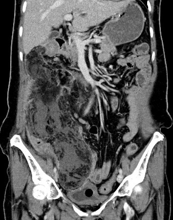
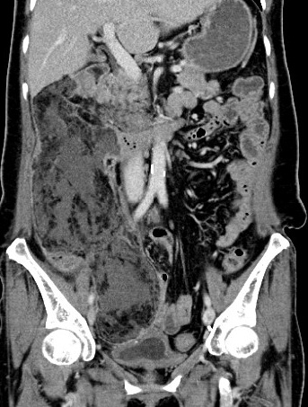
A 57-years-old woman who had a history of colonic carcinoma and recent laparoscopic right hemicolectomy, she presented with post-operative sepsis and leucocytosis. Plain and contrast CT abdomen and pelvis were performed.
Diagnosis:
Omental infarction
Discussion:
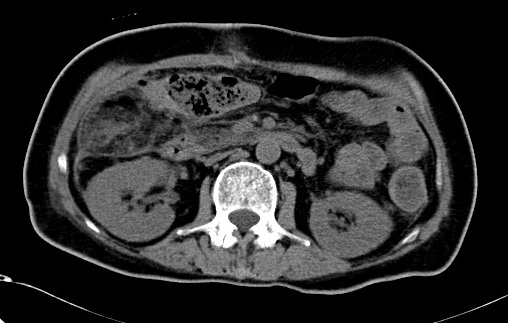
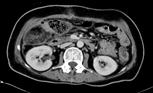
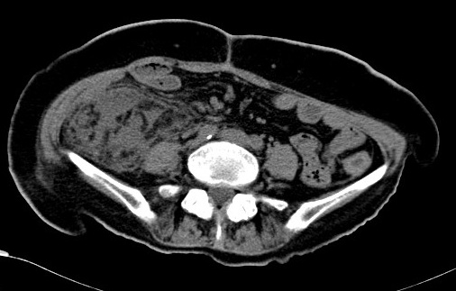
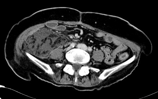
CT abdomen and pelvis showed a huge mass containing fat and soft tissue component in peritoneal cavity, extended from subhepatic region to right side of pelvic. The size measured ~5.4 x 7.9 x 27cm (APxWxH). The intralesional soft tissue component had low attenuation ( HU ~15 ) and did not show contrast enhancement.
Small bowel loops in right side abdomen was displaced laterally and compressed by the mass.
Peritoneal thickening along the right side abdomen adjacent to the mass was noted.
The preferred diagnosis is omental infarction. Given history of sepsis, superimposed infection/inflammation on the omental infarct should to be considered.
The typical radiographic features of omental infarct include nonenhancing fat-containing omental mass with heterogeneous attenuation, it lacks the hyperattenuating ring that is seen in epiploic appendagitis. The lesion in omental infarction is larger and most commonly is located in right-sided abdomen next to the cecum or the ascending colon, while epiploic appendagitis is most often less than 5 cm long and is located adjacent to the sigmoid colon.
Abnormal thickening and infiltration of the omentum has wide range differential diagnosis. Ranged from acute, focal, and diffuse infectious causes (appendicitis, diverticulitis, and peritonitis), to vascular causes (omental infarct and epiploic appendagitis), to chronic processes (carcinomatosis, mesothelioma, lymphoma, and tuberculosis).
In the acute clinical setting, infection and infarct of omentum should be considered. Although perfused by numerous small vessels, the omentum has a vascular supply that is relatively less redundant than that of the small or large bowel because fewer collaterals are present. Although the exact pathogenesis of acute omental infarction is not known, it is thought to be the result of an anomalous blood supply, particularly to the right lower quadrant of the omentum, with mechanical factors inducing venous thrombosis. Thus, as a general rule, omental infarcts occur in the right abdomen. Accepted predisposing factors include venous kinking due to increased abdominal pressure, compression of the omentum between the liver and the anterior abdominal wall, various causes of vascular congestion (postprandial causes, particularly in obese patients, coughing, Valsalva maneuver; and right-sided heart failure), and recent surgery.
In conclusion, omental infarction is a rare, self-limited process, with conservative management, the patient's symptoms usually resolve within 2 weeks.
