Answer of December 2014
For completion of the online quiz, please visit the HKAM iCMECPD website: http://www.icmecpd.hk/
Clinical History:
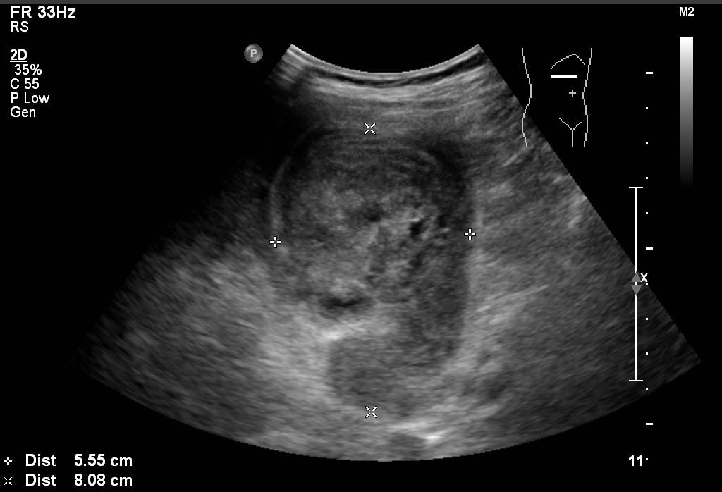
A 70-year-old gentleman is admitted with fever and upper abdominal pain for 3 days. He is febrile and physical examination revealed right upper quadrant tenderness. Initial blood workup showed elevated white cell count with deranged liver function.
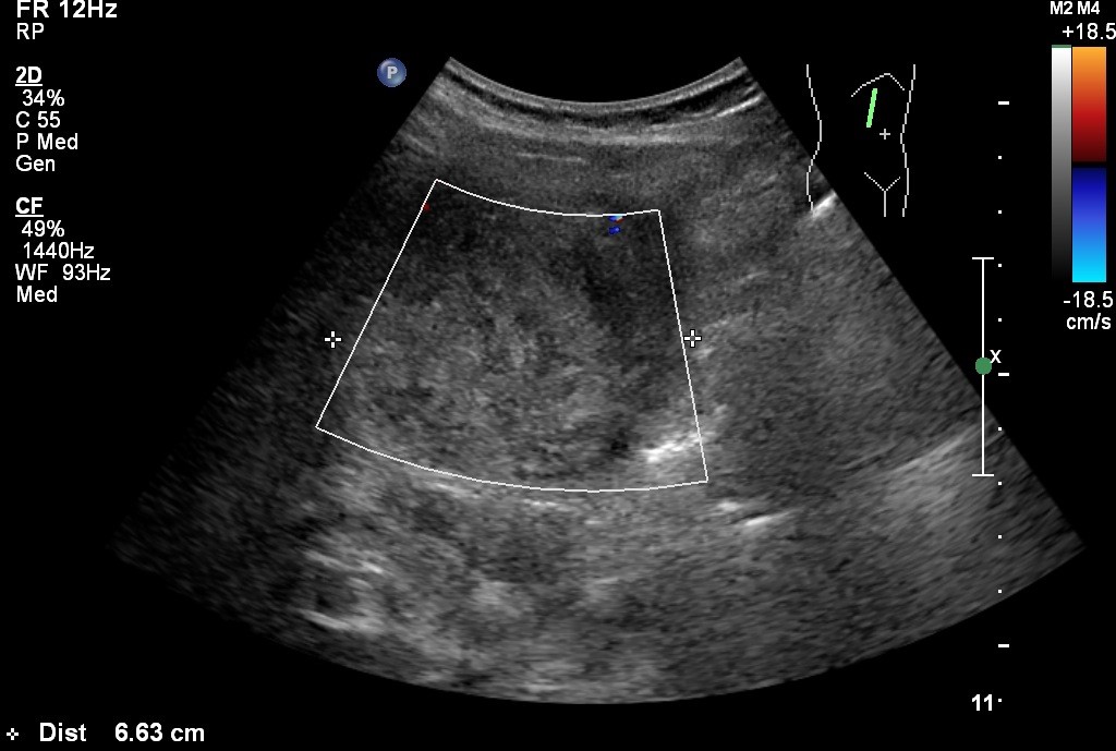
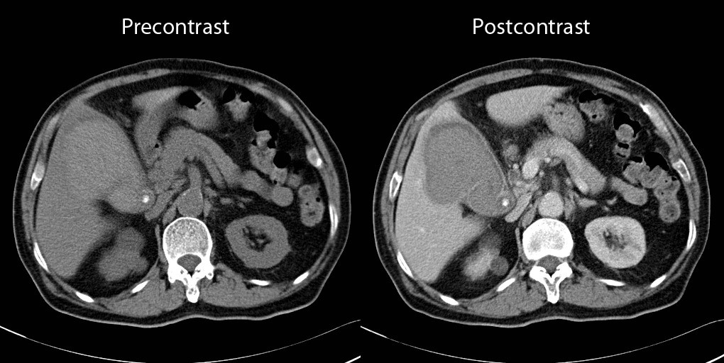
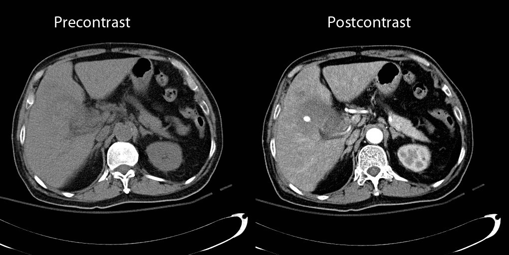
Ultrasound abdomen and later CT abdomen with contrast are obtained.
Diagnosis:
Discussion:
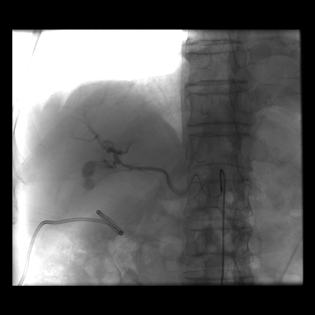
Hemorrhagic cholecystitis is a rare complication of biliary tract disease but potentially fatal. They can occur in the setting of trauma, malignancy and in patient with bleeding tendencies (such as renal failure, cirrhosis and anticoagulation). Patients usually present with signs and symptoms of acute calculous cholecystitis, like right upper quadrant pain, fever and leukocytosis. However, as they have hemorrhage into the gallbladder, they may also have other symptoms like hemobilia, melena or hematemesis. Ultrasound may be able to demonstrate intracholecystic echogenic mass-like lesions without posterior acoustic shadowing. Color Doppler can be used to confirm the absence of blood flow within the mass, making the diagnosis of gallbladder tumor less likely. On CT scan, precontrast hyperdense non-enhancing gallbladder contents representing acute blood product can be seen. On post contrast images, hyperdense focus representing pseudoaneurysm or active contrast extravasation may be detected. Treatment options should consist of emergency cholecystectomy with ligation of cystic artery. But in those with high surgical risks and acute hemorrhage, endovascular emoblization can be utilized with the aim of halting active hemorrhage into the biliary/gastrointestinal system or peritoneum.
