Answer of August 2014
Clincial History:
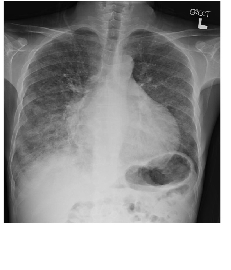
A 30 years old CAPD patient presented with shortness of breath. ( 9 images: chest X ray X1, HRCT axial X 5, Bone scan X 3)


Diagnosis:
Metastatic pulmonary calcification.
Discussion:
Metastatic calcification refers to deposition of calcium in normal tissue, such as lung, stomach, kidneys and heart. HRCT is more sensitive than plain radiograph in detecting calcium deposition. Imaging findings vary from multiple centrilobular mulberry-shaped amorphous calcifcation of 3mm to 10mm in diameter to non specific ground glass opacities and consolidation. In our case, there are multiple ground glass nodules and ground glass opacities seen in HRCT. Subsequent bone scan reveals intense abnormal uptake in the lungs, compatible with metastatic pulmonary calcification.
Patient can be asymptomatic or present with progresssive respiratory failure. In severe cases, patients can suffer from restrictive pulmonary function with decreased carbon monoxide diffusion. Cardiac involvement is possible. Death is usually resulted from involvement of conducting pathyways. Imaging findings may be reversible with correction of underlying hypercalcemia.
Plain film radiography and HRCT can to use to detect the amount of calcium depostition in tissues, while bone scintigraphy can depicts the active process of calcium phosphate metabloism.