Answer of May 2021
For completion of the online quiz, please visit the HKAM iCMECPD website: http://www.icmecpd.hk/
Clinical History:
A 70 years old female patient presents with neck discomfort
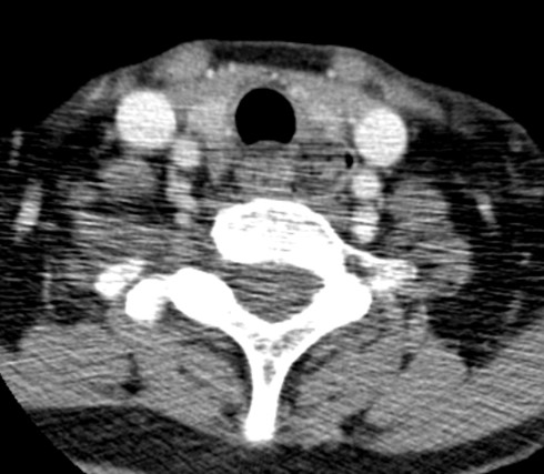
Axial image of contrast CT neck
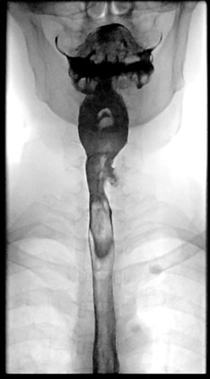
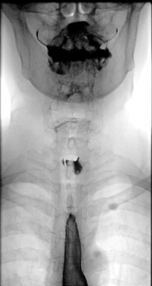
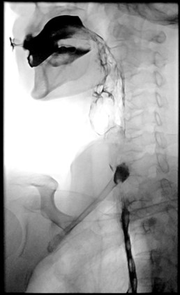
Selected images of barium swallow (frontal and lateral views)
IMAGING FINDINGS:
Axial image of contrast CT neck shows a lesion on the left side of the upper oesophagus with internal mottled gas densities.
DIAGNOSIS:
Killian-Jamieson diverticulum
DISCUSSION:
Killian-Jamieson diverticulum is an uncommon diverticulum which arises from the upper oesophageal region. Anatomically, it is a protrusion through a congenital weakness point named Killian-Jamieson space (not to be confused with Killian’s dehiscence). This space is bounded superiorly by the cricopharyngeus muscle, anteriorly by the posterior wall of the cricoid cartilage, and inferomedially by the longitudinal tendon of the oesophagus as it inserts onto the cricoid cartilage. It is where the recurrent laryngeal nerve enters the pharynx
Zenker’s diverticulum is another type of diverticulum that also arises from the upper oesophageal region. It originates at the posterior wall of the pharyngoesophageal segment in a midline area of weakness just above the cricopharyngeus (Killian's dehiscence).
Killian-Jamieson diverticula are four times less frequent than Zenker’s diverticula. They tend to be smaller than Zenker’s diverticula. They occur more frequently on the left side but can be bilateral. Killian-Jamieson diverticula are less likely to cause symptoms and are less likely to be associated with overflow aspiration or gastroesophageal reflux than Zenker's diverticula.
On barium swallow, the opening of a Killian-Jamieson diverticulum is located just below the level of the cricopharyngeus, with the sac of the diverticulum lying lateral to the cervical oesophagus on frontal images and overlapping the anterior wall of the cervical oesophagus on lateral images. On the contrary, the opening of a Zenker's diverticulum is above the cricopharyngeus, with the sac of the diverticulum lying posterior to the cervical oesophagus on lateral images and in the midline on frontal images.