Answer of November 2018
For completion of the online quiz, please visit the HKAM iCMECPD website: http://www.icmecpd.hk/
Clinical History:
This 42-year-old lady complained of progressive enlargement of right arm mass. Clinically the mass was tender with irregular contour. No superficial skin discoloration nor lesion detected.
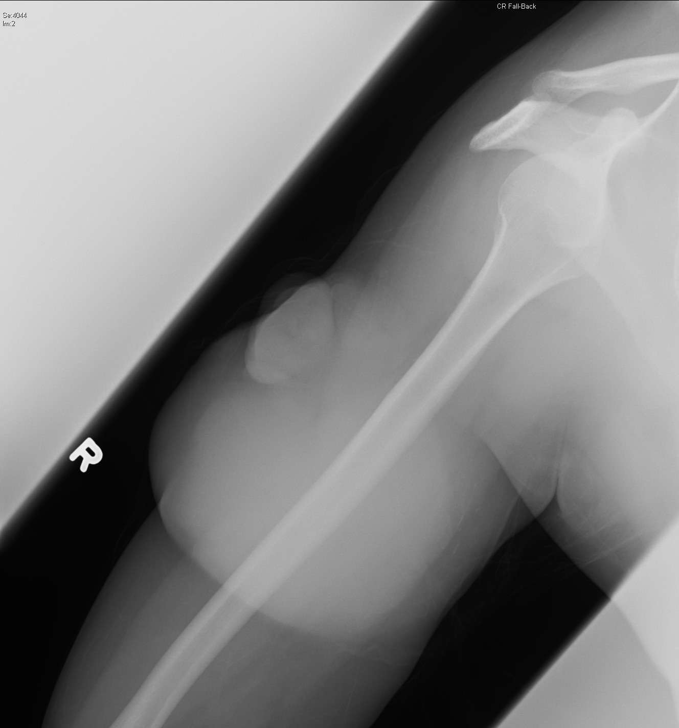
Right Arm Radiograph
Image 1 of 4
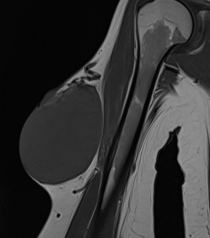
Right Arm MRI (T1)
Image 2 of 4
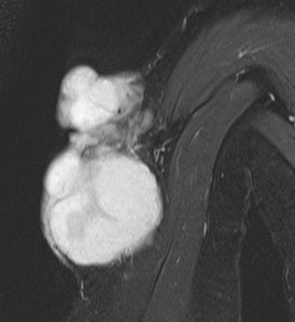
Right Arm MRI (T2)
Image 3 of 4
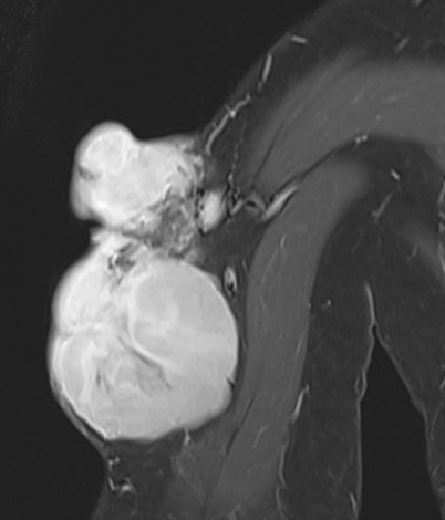
Right Arm MRI (T1 with contrast)
Image 4 of 4
Imaging Findings:
- Right Shoulder Radiograph - Large soft tissue swelling at the mid humeral shaft region noted without underlying bone destruction.
- MRI Right Arm - Large fungating but encapsulated tumour centered at subcutaneous layer with no invasion of deep fascia. It is isointense to muscle in T1 and hyperintense in T2, with avid enhancement. Enlarged tortuous feeding and draining vessels.
Diagnosis:
Dermatofibrosarcoma protuberans of right upper arm.
Discussion:
Dermatofibrosarcoma protuberans is a cutaneous soft tissue sarcoma involving the dermis layer. 85-90% of cases are low grade, only 2-5% of cases with high grade component.
It is a rare tumour with estimated incidence around 1 per million people per year. It is usually diagnosed in the 30s but found in all age groups.
More than 90% of cases have unique translocation t(17;22) which leads to self stimulatory growth into tumor.
Patients usually present with asymptomatic indurated plaque, which on palpation is a firm nodule fixed to dermis but move freely over deeper muscles. It slowly enlarges over months to years. Early lesions may be violet-red or blue, surrounding skin may be telangiectatic. It is usually located at the trunk or shoulders.
Classical imaging characteristics include its subcutaneous location with nodular exophytic pattern. A clean flat plane usually separates it from muscle and deep fascia. On cross sectional imaging, its CT density / MR intensity and enhancement are non-specific.
For large cutaneous / subcutaneous malignant appearing mass, imaging appearance is specific for dermatofibrosarcoma protuberans.
For less aggressive appearing and smaller lesions, the list of differentials is long.
|
Entity |
Features of this entity |
Features of dermatofibrosarcoma protuberans |
|
Dermatofibroma |
Commonly in extremities |
more commonly in the trunk |
|
Epidermal cyst |
No contrast enhancement unless infected |
variable enhancement |
|
Peripheral nerve sheath tumour |
Close to major nerve (split fat / fascicular / target sign) |
No particular relationship to nerve |
|
Nodular fasciitis |
Fascial tail sign (linear extension of lesion along fascia) |
|
|
Cavernous hemangioma |
Low density on CT +/- calcification |
|
|
Liposarcoma |
contains fat |
|
|
Nodular melanoma |
Clinically blue to black |
Clinically violaceous |
|
Melanocytic melanoma |
MR T1 hyperintense, T2 hypointense +/- bone / lymph node metastases |
|
CT or MRI is helpful in delineating local extent and exclude bone involvement. Dermatofibrosarcoma protuberans rarely metastasizes. If it does, it more likely involve the lungs rather than lymph nodes.
Diagnosis is by core or excisional biopsy of suspected lesions, with histological and immunohistochemistry assessment. Molecular testing is done if diagnosis is in doubt.
Treatment is by surgical resection, with or without adjuvant radiotherapy and chemotherapy.
Post-operative outcome is usually good, although local recurrence is common.