Answer of August 2017
For completion of the online quiz, please visit the HKAM iCMECPD website: http://www.icmecpd.hk/
Clinical History:
Middle aged waitress with relative good past health presented with slow growing mass over the left forearm for 2 years. She also complained of mild numbness along the median nerve territory. Physical examination reveals a firm mobile mass over the forearm at the volar aspect, measuring at 15cm x 10cm.
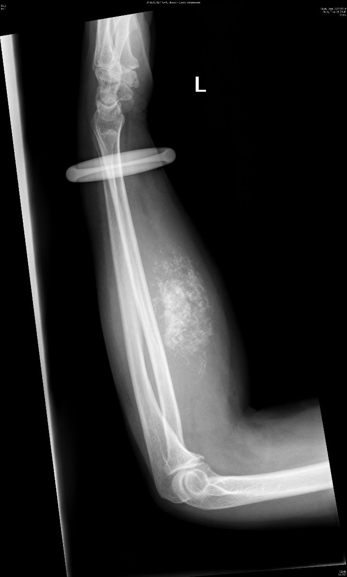
Figure 1. Plain XR Left Forearm (Lateral)
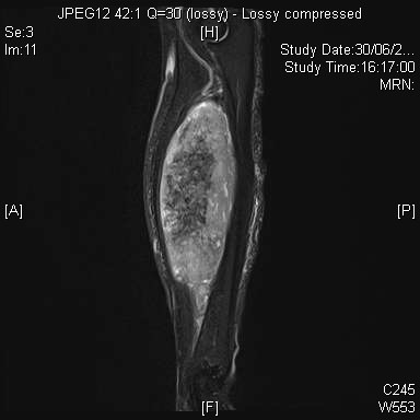
Figure 2. STIR
Imaging Findings:
Plain Radiograph shows the presence of soft tissue swelling over the left forearm. It is associated with ring-and-arc calcifications. No bony involvement noted. The underlying bone (i.e. the radius and ulnar) shows no abnormal periosteal reaction.
Diagnosis:
Extra-skeletal chondrosarcoma (Mesenchymal Type)
Discussion:
Extra-skeletal chondrosarcoma is a rare type of soft tissue sarcoma. It only accounts for 1% of all chondrosarcomas and tends to be of a higher grade than conventional type of intramedullary chondrosarcoma. It is further subdivided into myxoid or mesenchymal type according to its pathology.
The myxoid type chondrosarcoma usually affects the middle age and commonly involves the thigh, while the mesenchymal type is seen more in younger adults at the head and neck region.
Mesenchymal chondrosarcoma was first described in 1959. It is a rare specific subtype with slight female predominance. Various case reports have been published with the brain, meninges, orbits and muscles of lower limbs being involved. Main differential diagnosis includes synovial sarcoma, Ewings and hemangiopericytoma.
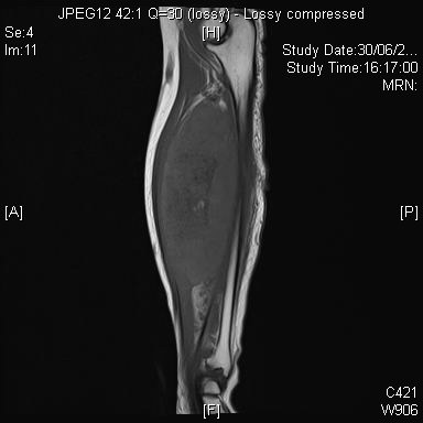
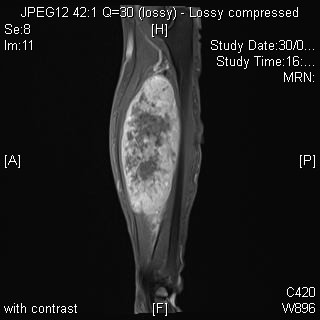
It has a characteristic pathological pattern with a Bimorphic appearance. It has both the small blue cell component, as seen in Ewings, neuroblastoma and PNET and also the cartilaginous component. Due to such a characteristic pattern, its MRI findings is less typical than those we see in chondroid tumours. Less water content is expected due to the mixture of small blue cells. MRI usually shows an intermediate T2 signal unless there is presence of necrosis. It also shows diffuse heterogenous enhancement after administration of contrast. Due to its non-specific MRI findings, it is important to look for chondroid calcifications/rings and arcs calcifications in plain radiographs as it may be the only clue to diagnosis, as shown by the case above.


