Answer of August 2016
For completion of the online quiz, please visit the HKAM iCMECPD website: http://www.icmecpd.hk/
Clinical History:
An 80-year-old elderly gentleman with chronic right shoulder pain presents with a progressively enlarging mass in right upper shoulder over a few months.
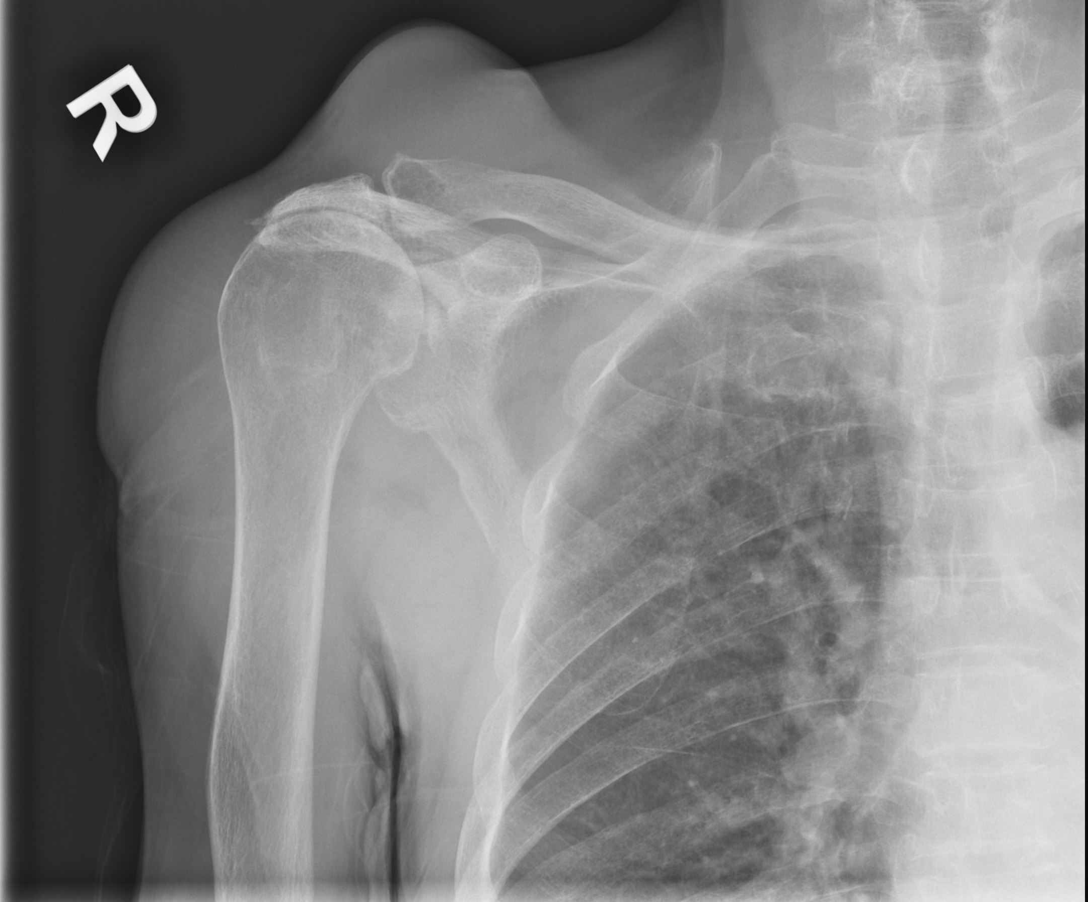
Frontal right shoulder radiograph
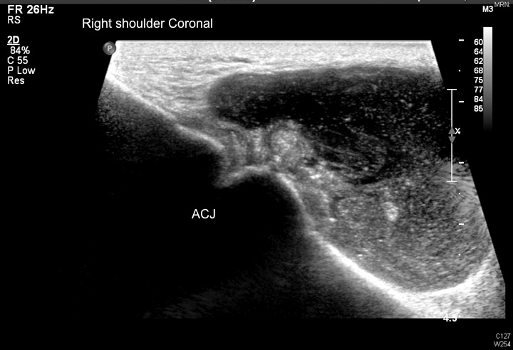
Coronal ultrasound at Acromioclavicular joint
Diagnosis:
Acromioclavicular joint cyst.
Backgound rotator cuff arthropathy.
Discussion:
The frontal radiograph shows a large mass of soft tissue density superior to the degenerative right acromioclavicular joint. There is marked narrowing of the subacromial space with superior migration of humeral head, which shows ‘femoralization’, and ‘acetabulization’ of the coracoacromial arch.
Ultrasound confirms the presence of a unilocular cystic mass with tapering towards the acromioclavicular joint. Internal debris is present. Massive rotator cuff tear is confirmed on other images (not shown).
Majority of reported cases of acromioclaivuclar joint cysts are associated with rotator cuff pathology, as in our case. With rotator cuff insufficiency, pull of deltoid causes superior migration of humeral head, striking and tearing the inferior and then superior portion of the acromioclavicular joint capsule. Over time the defect allows glenohumeral synovial fluid to decompress across the acromioclaivulcar joint and escape superiorly to the subcutaneous tissue in the form of a synovial cyst. Uncommonly this could occur in an intact cuff. Detection of tear is essential for planning of subsequent treatment.
The loss of stabilizing function of the rotator cuff can lead to a complex pattern of joint degeneration referred to as rotator cuff arthropathy, characterized by massive rotator cuff tear, degenerative and reciprocal changes.
Traditionally, these cysts were identified with shoulder arthrography as a "geyser" of fluid escaping through the acromioclavicular joint. With increasing use of MRI, a MR-equivalent ‘geyser sign’ has been described with synovial fluid through the cuff defect, across subacromial bursa and decompressing superiorly through a degenerated acromioclavicular joint.
Treatment includes expectant management or surgical intervention, including arthrodesis, joint debridement and arthroplasty depending on patients’ functional demand and co-morbidities.

