Answer of October 2015
For completion of the online quiz, please visit the HKAM iCMECPD website: http://www.icmecpd.hk/
Clinical History:
A 50-year old woman who presented with post-menopausal bleeding was incidentally found to have a bladder mass on transvaginal ultrasound. Cystoscopy showed an irregular mucosal mass at the dome of the urinary bladder. Biopsy of the mass revealed adenocarcinoma, thus CT was performed to look for evidence of colonic malignancy with invasion to the bladder.
Discussion:
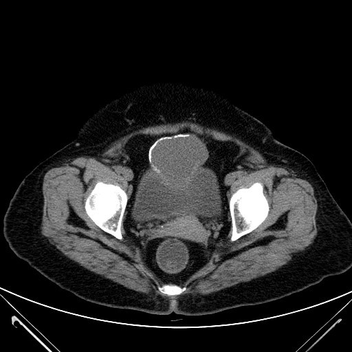
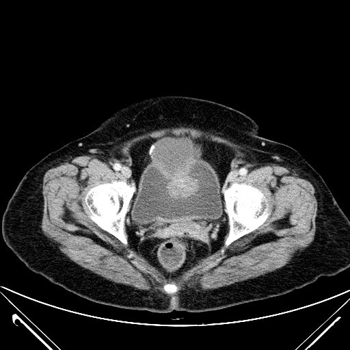
Non-contrast and post-contrast CT of the pelvis demonstrates a partially intravesical and partially extravesical mass located in the midline at the anterior bladder dome. It is of mixed attenuation with both low attenuation and enhacing soft tissue attenuation components. Curvilinear peripheral calcification is noted around the low attenuation component. This lesion is separate from adjacent bowel loops. Location of the mass and its features are characteristic of a urachal remnant tumour.
The urachus (median umbilical ligament) is a vestigial remnant of the cloaca (fetal bladder precursor) and allantois (yolk sac), which normally involutes before birth, remaining as a fibrous band in adulthood. It extends from the bladder to the umbilicus in the preperitoneal perivesical space; occasionally it may slightly deviate to the right or left of midline if it merges with one or the obliterated umbilical arteries. It consists usually of transitional epithelium (70%).
Urachal neoplasms are rare. Benign neoplasms are extremely rare, and their significance lies in that they may mimick malignant neoplasms. Malignant urachal neoplasm (urachal carcinoma) predominantly manifests as adenocarcinoma (90%), probably due to metaplasia of urachal mucosa into columnar epithelium followed by malignant transformation. Adenocarcinomas arising from the urinary bladder may be primary, urachal in origin or metastatic: 34% are of urachal origin. At histological analysis, mucin production is found in up to 75% of cases, which accounts for a characteristic imaging finding of low-attenuation components within the tumour. 90% of urachal carcinomas arise in the juxtavesical portion of the urachus. They may be confused with primary tumours of the bladder dome, but urachal tumours tend to grow in the perivesical space toward the umbilicus.
Characteristic findings of this tumour includes the presence of an unencapsulated caudal part involving a portion of the bladder wall and an often cystic encapsulated supravesical portion. Calcification occurs in 50-70% and pay be punctate, stippled, or curvilinear and peripheral, which is due to histological finding of typical psammomatous calcifications as with some other mucinous adenocarcinomas. It may occasionally be difficult to distinguish an infected urachal remnant from urachal carcinoma. Hematuria, mural nodularity, calcification and CT and lack of adjacent inflammatory change point towards the latter diagnosis.
The commonest presenting symptom is hematuria. Prognosis in urachal tumours is generally poor as the tumour arises in a clinically silent area and is discovered only after it has grown to a large size or extended into the bladder lumen or adjacent organs. Metastases occur in the pelvic lymph nodes initially, followed by systemic metastases to lung, brain, liver, bone.

