Answer of September 2015
For completion of the online quiz, please visit the HKAM iCMECPD website: http://www.icmecpd.hk/
Clinical History:
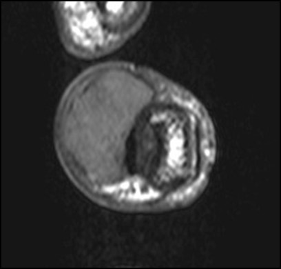
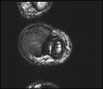
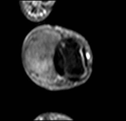
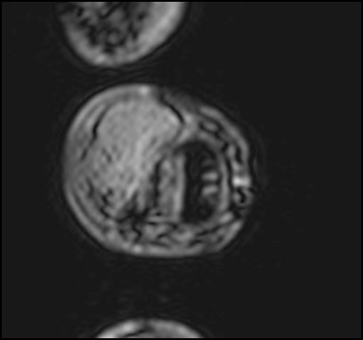
A 66 year-old gentleman presented with left middle finger swelling for 2 years. It was slowly increasing in size, with no pain or numbness. Physical examination showed a firm multi-lobulated mass of about 1.5cm over the volar aspect of left middle finger at the distal interphalangeal joint level. Magnetic resonance imaging (MRI) was performed (4 images – upper row: axial T1-weighted image, axial T2-weighted image; lower row: post-contrast fat-suppressed axial T1-weighted image, axial T2-weighted gradient-echo image)
MRI
Diagnosis:
Discussion:
MRI is useful in the pre-operative diagnosis of giant cell tumor of tendon sheath. MR imaging features of giant cell tumor of tendon sheath reflect its underlying histological characteristics. On T1-weighted and T2-weighted images, it has low or intermediate signal intensity, due to the presence of hemosiderin deposition within the lesion, which exerts paramagnetic effect that shortens T1 and T2 relaxation times. The paramagnetic effect of hemosiderin is exaggerated on gradient-echo sequences due to increased magnetic susceptibility. Contrast enhancement is due to the presence of numerous proliferative capillaries in the collagenous stroma.
The major differential consideration for giant cell tumor of tendon sheath in the hand is fibroma of the tendon sheath. It can be confused with giant cell tumor of tendon sheath clinically, radiologically and even pathologically. In MR imaging, it usually appears as hypointense to isointense on both T1-weighted and T2-weighted sequences with no or little enhancement due to abundant acellular fibrous tissue with areas of hyalinization. However, it may have areas of high T2 signal or enhancement, depending on their compositions of fibrous tissue, myxoid change and cellularity.
Local surgical resection is the mainstay of treatment for giant cell tumor of tendon sheath, with recurrence rate seen in 7-44% of cases.


