Answer of November 1998
Clinical History:
F/26. Vomiting and acute confusion for 2 weeks. GCS 10/15 on admission. Died after 1 week.
Figure 1 CT brain
Figure 2 Contrast enhanced CT brain
Figure 3 CXR
What is your diagnosis?
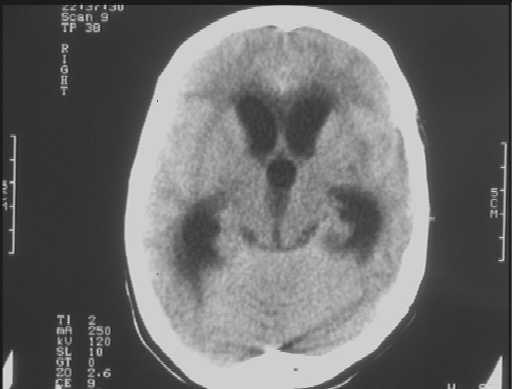
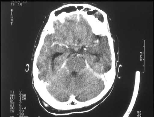
Figure 1 Figure 2

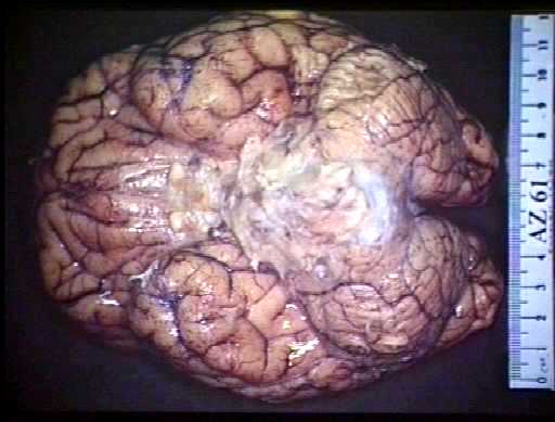
Figure 3 Figure 4
Diagnosis:
TUBERCULOUS MENINGITIS
Discussion:
Figure 1 CT showed hydrocephalus.
Figure 2 CECT basal enhancement not prominent.
Figure 3 CXR showed consolidation in posterior basal segment of left lower lobe.
Figure 4 Post-mortem showed TB meningitis with endarteritis and thrombosis, abscess in left lower lobe and miliary TB in lung.