Answer of September 1999
Clinical History:
F/78 Chest pain
Figure 1 CXR(PA)
Figure 2 CXR
Figure 3 CXR(lateral)
What is your diagnosis?
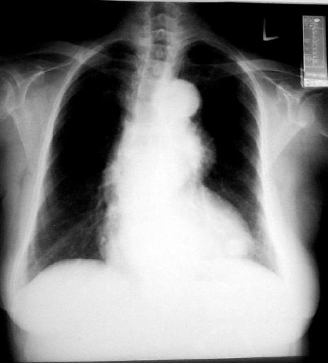
Figure 1
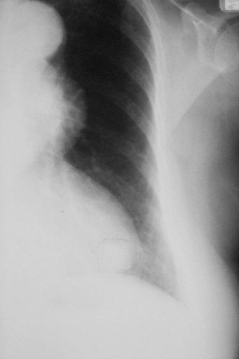
Figure 2
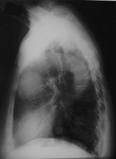
Figure 3
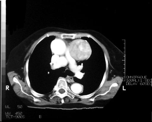
Figure 4

Figure 5
Diagnosis:
Malignant thymoma
Discussion:
Figure 1 CXR(PA) shows a well defined mass in the anterior mediastinum and a small retrocardiac lesion .
Figure 2 CXR localised view better demonstrates both the anterior mediastinal mass and retrocardiac lesion.
Figure 3 CXR (lateral) just comfirmed the location of both masses to a better advantage.
Figure 4 CECT thorax shows a heterogenous enhancing mass in the anterior mediastinum.
Figure 5 CECT thorax shows the retrocardiac mass is actually two masses abutting the dome of left hemidiaphragm. Biopsy revealed malignant thymoma.