Answer of January 2000
Clinical History:
F/67 Rountine follow up in cardiac clinic
Figure 1 CXR
What is your diagnosis?
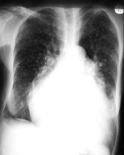
Figure 1
Diagnosis:
Pulmonary calcifications in mitral valve disease.
Discussion:
Figure 1 CXR shows bilateral multiple small pulmonary calcified nodules predominantly in the mid and lower zones. There is also evidence of mitral valve heart disease with cardiomegaly, enlarged left atrium , widening of carina and pulmonary venous hypertension.