Answer of August 2000
Clinical History:
F/15 with sudden onset of headache.
Figure 1 (NECT)
Figure 2 (NECT)
Figure 3 (T1WI)
Figure 4 (T1WI, post GD)
What is your diagnosis?

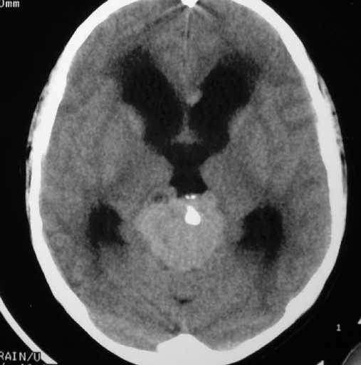
Figure 1 Figure 2
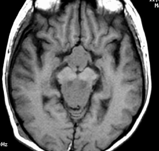
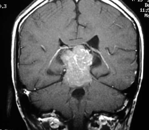
Figure 3 Figure 4
Diagnosis:
Pineal and Suprasellar Germinomas
Discussion:
Figure 1&2 CT shows 2 hyperdense masses in the suprasellar and pineal regions. The latter contains calcifications and cystic areas. There is also obstructive hydrocephalus.
Figure 3&4 MR shows the masses are hypointense on T1WI and are very enhancing.