Answer of January 2001
Clinical History:
F/13 with dysphagia
Figure 1 (CECT)
Figure 2 (CECT)
What is your diagnosis?
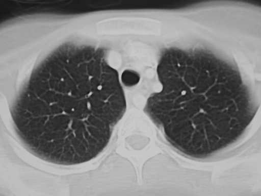

Figure 1 Figure 2
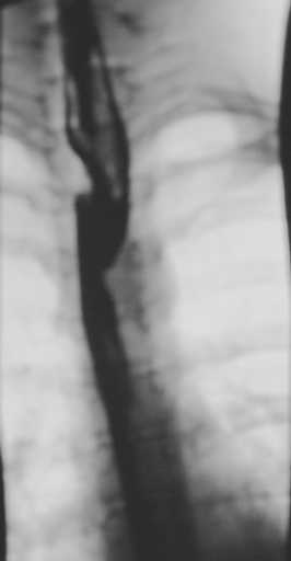
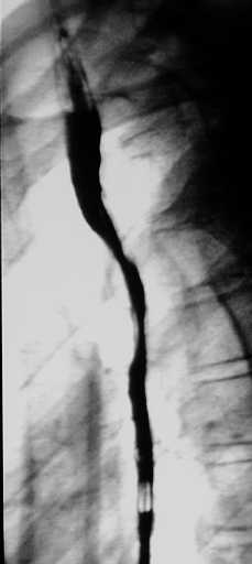
Figure 3 Figure 4
Diagnosis:
Aberrant right subclavian artery
Discussion:
Figure 1 & 2 CT shows an aberrant vessel behind the esophagus from the left running upward to the right.
Figure 3(AP)& 4(Lateral) Barium swallow demonstrates the characteristics oblique and posterior indentations respectively.