Answer of February 2001
Clinical History:
renal failure patient admitted for hyponatremia and hyperkalemia. Initial CT brain is normal. Condition deteriorates 1 week later. MR performed 3 weeks later.
Figure 1 (T1WI)
Figure 2 (T2WI)
Figure 3 (T2WI)
What is your diagnosis?
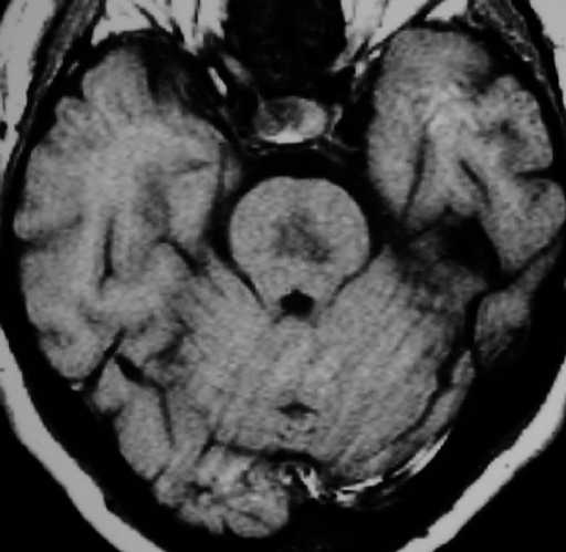
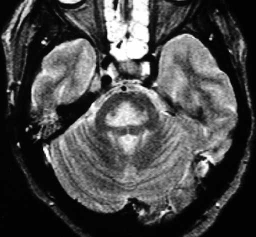
Figure 1 Figure 2
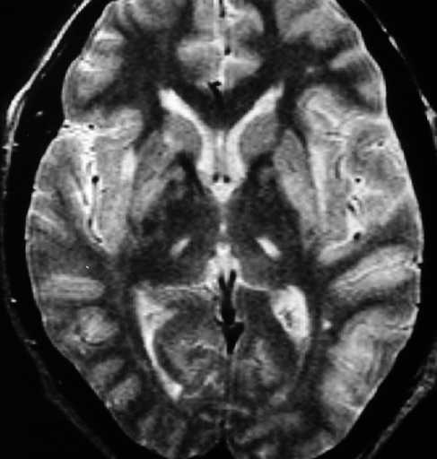
Figure 3
Diagnosis:
Osmotic Myelinolysis (central pontine myelinolysis & extrapontine myelinolysis)
Discussion:
Figure 1 & 2 MR shows a large T1 hypo & T2 hyperintense lesion in the central pon.
Figure 3 There are also T2 hyperintense lesions in the bilateral basal ganglia and thalami.