Answer of October 2002
Clinical History:
M/18, cough with blood stained sputum for 2 weeks.
Figure 1: Frontal chest radiograph.
Figure 2: Noncontrast axial CT scan of thorax.
Figure 3: Contrast axial CT scan of thorax.
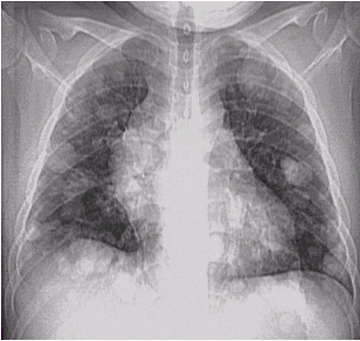
Figure 1
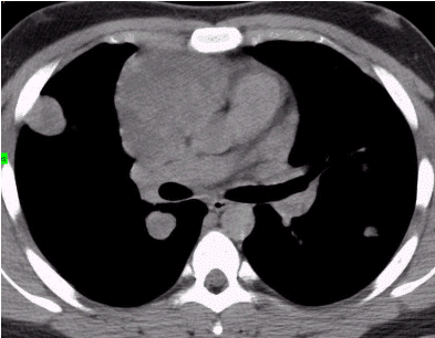
Figure 2
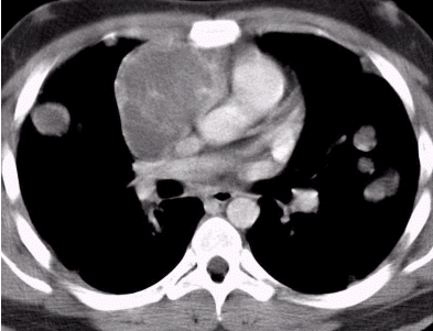
Figure 3
Diagnosis:
Choriocarcinoma of mediastinum with lung metastases.
Discussion:
Figure 1: Frontal chest radiograph shows mediastinal widening with multiple "cannon ball" lesions in both lungs, more in the lower zones.
Figure 2 and 3: CT thorax shows an non-calcified solid anterior mediastinal mass slightly hypodense to the surrounding medistinal structure and with heterogeneous enhancement. Multiple nodules are seen in both lungs.