Answer of November 2003
Clinical History:
F/25. Abdominal discomfort.
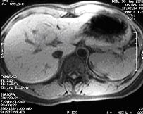
Precontrast T1-weighted
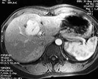
Postcontrast Arterial phase T1-weighted
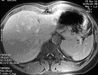
Postcontrast Delayed phase T1-weighted
Diagnosis:
Pulmonary varix t
Discussion:
- A 3 cm well-defined right paracardiac opacity is seen on the frontal CXR. The opacity is situated in the right lower lobe, as it is posterior to the major fissure on the lateral view.
- The lesion is demonstrated as a dilated right inferior pulmonary vein, draining into the left atrium. It shows comparable contrast enhancement to the left atrium.