Answer of June 2005
Clinical History:
46-year-old female with recent abdominal discomfort.
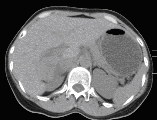
CT abdomen (plain)
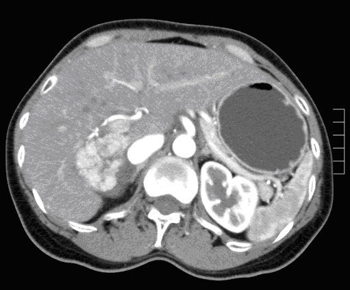
CT abdomen (arterial phase)
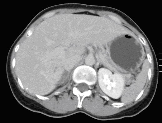
CT abdomen (portovenous phase)
CT abdomen (delayed 5 minutes)
Diagnosis:
Focal Nodular Hyperplasia
Discussion:
A large well defined noncalcified isodense mass is noted in the right lobe of liver measuring about 6cm. The lesion shows intense enhancement in the arterial phase and becomes isodense to liver parenchyma in the portovenous and delay scans. Small central triangular hypodensity is noted, suggesting scar. The diagnois is focal nodular hyperplasia. Previous scans show no significant change with follow up studies.


