Answer of November 2005
Clinical History:
62-year-old lady presented with one day history of dizziness and elevated blood pressure. She had history of chronic renal failure and diabetes mellitus. After hospital admission, she developed status epilepticus.


Diagnosis:
Posterior reversible encephalopathy syndrome
Discussion:
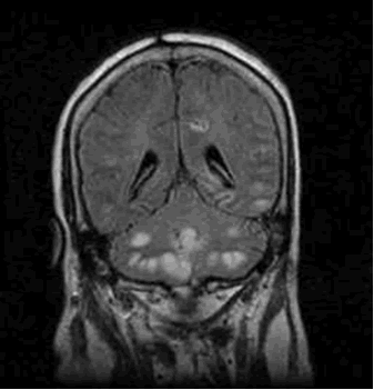
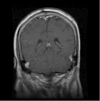
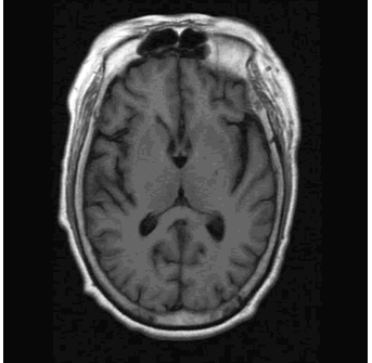
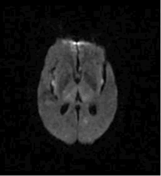
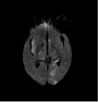
MRI (T1W, T2W, FLAIR, DWI) showed multiple cortical and subcortically lesions in bilateral cerebellar hemispheres, left side of pons, bilateral thalami and bilateral insular regions, left para-hippocampus and bilateral cerebral cortex. They predominantly distribute over the cerebellum and occipital region. They are hypointense on T1WI images and hyperintense on T2WI and FLAIR images and showed restricted diffusion on diffusion-weighted study. There was mild mass effect and no contrast enhancement.
The distribution of lesions in posterior reversible encephalopathy syndrome (PRES) is almost always bilateral and rather symmetrical. They involve primarily the white matter, but cortex is also involved. Typical site involves posterior portions of both cerebral hemispheres, especially the parieto-occipital regions. Other posterior structures, such as cerebellum and brainstem, are also commonly affected. Involvement of the deep gray nuclei can also occur. Contrast enhancement is variable. Although the lesions predominate in posterior circulation territories, lesions of the anterior circulation are also seen. Anterior abnormalities tend to be seen only in the most severe cases and are usually associated with edema in the posterior circulation territories.