Answer of January 2006
Clinical History:
A 68 year old gentleman who developed sudden onset of dysarthria and dizziness during a prolonged dental procedure. When he was taken to the emergency room, he was unconscious and required intubation. The following examinations were done 2 days after onset of symptoms.


Plain CT brain Plain CT brain
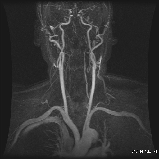

CE-MRA of the neck vessels D TOF MRA of circle of Willis


SE T1W EPI-DWI
Diagnosis:
Vertebral artery dissection
Discussion:
The imaging findings are that of steno-occlusive pattern of vertebral artery dissection. The contrast enhanced MR anigogram clearly shows the tapered occlusion of the vertebral arteries at V3 levels. V2 segments also show irregular filling defects. Very little reconstitution of intracranial vertebral and basilar arteries can be seen. Also noted the high signal in the left intracranial vertebral artery on T1W axial MR image, probably due to subacute clot inside or very slow flow. T1W axial scan of the neck failed due to patient movement despite sedation. The classical finding would be a crescent of high signal intramural haematoma.