Answer of August 2006
Clinical History:
A 70-years-old lady with history of arrhythmia presented with shortness of breath. HRCT of the thorax was obtained.


Diagnosis:
Amiodarone-induced lung fibrosis
Discussion:
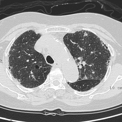
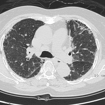
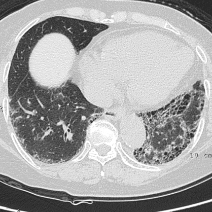
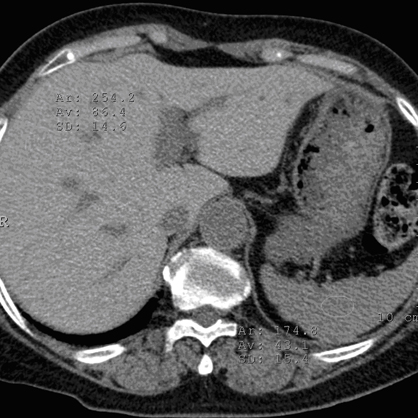
HRCT thorax showed architectural distortion with interlobular septal thickening and traction bronchiectasis in both lungs, predominantly basal and peripheral in distribution. It is most severe in left lung base with honeycomb appearance. Findings are compatible with pulmonary fibrosis. Cardiomegaly was an additional finding. Nonenhanced abdominal CT scan showed high attenuation of the liver (86HU) relative to that of the spleen (43HU), indicating abnormally hyperdense hepatic parenchyma. The patient was actually receiving amiodarone for cardiomyopathy and arrhythmia and the overall picture is compatible with amiodarone-induced lung fibrosis and hyperdense liver.