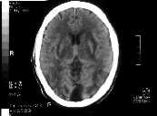
Figure 1
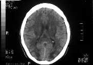
Figure 2
Figure 1 NECT brain shows bilateral symmetrical hypodensities in internal and external capsules, globus pallidus and mainly the subcortical white matter of the occipital lobe.Figure 2 NECT brain better demonstrates the occipital lobe hypodensities predominantly affecting the subcortical white matter.This patient has both uraemic (basal ganglia changes) and hypertensive (occipital lobe changes) encephalopathy.
| ||||
HISTORY |
PREVIOUS MTHS |
|||
HOME |
CURRENT MTH |
|||