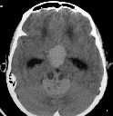
Figure 1
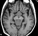
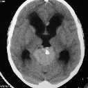
Figure 2
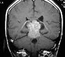
Figure 1&2 CT shows 2 hyperdense masses in the suprasellar and pineal regions. The latter contains calcifications and cystic areas. There is also obstructive hydrocephalus.Figure 3&4 MR shows the masses are hypointense on T1WI and are very enhancing.
| ||||
HISTORY |
PREVIOUS MTHS |
|||
HOME |
CURRENT MTH |
|||