Figure 1
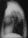
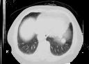
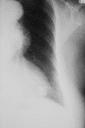
Figure 2

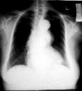
Figure 1 CXR(PA) shows a well defined mass in the anterior mediastinum and a small retrocardiac lesion .Figure 2 CXR localised view better demonstrates both the anterior mediastinal mass and retrocardiac lesion.Figure 3 CXR (lateral) just comfirmed the location of both masses to a better advantage.Figure 4 CECT thorax shows a heterogenous enhancing mass in the anterior mediastinum.Figure 5 CECT thorax shows the retrocardiac mass is actually two masses abutting the dome of left hemidiaphragm. Biopsy revealed malignant thymoma.
| ||||
HISTORY |
PREVIOUS MTHS |
|||
HOME |
CURRENT MTH |
|||