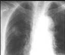
Figure 1
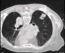
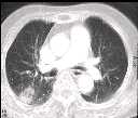
Figure 2
Figure 1 CXR (PA) showed R mid zone soft opacity.Figure 2 CECT showed peripheral lung lesion in posterior segment of R upper lobe. No lymphadenopathy.Figure 3 CT guided biopsy in prone position.
| ||||
HISTORY |
PREVIOUS MTHS |
|||
HOME |
CURRENT MTH |
|||