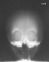
Figure 1
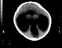
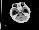
Figure 2
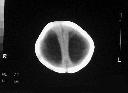
Figure 1 SXR shows midline bony defect in the maxilla and hypoteloric orbits.Figure 2 NECT brain shows dilated bilateral temporal horns and normal posterior fossa structure.Figure 3 NECT brain shows a dorsal cyst communicating with partiarlly formed lateral ventricles. The thalami are partially fused across the midline with absent septum pellucidum.Figure 4 NECT brain demonstrates present of falx cerebrii and more distinct seperation of both cerebral hemisphere. This baby has semiobar holoprosencephaly.
| ||||
HISTORY |
PREVIOUS MTHS |
|||
HOME |
CURRENT MTH |
|||