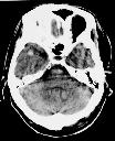
Figure 1
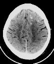
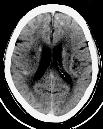
Figure 2
Figure 1 NECT brain shows small hyperdense focus in the right temporal pole and left upper pons of the brain.Figure 2 NECT brain show further multiple hyperdense foci in the grey-white matter junction of left frontal and right parietal lobes. Incidentally bilateral old lacunar infarcts in the corona radiata are seen.Figure 2 NECT brain again demontrates the bilateral grey- white matter multiple small haemorrhages at the classic parasaggital region of both frontal lobes.These findings are characteristic of diffuse axonal injury (white matter shearing injury.)
| ||||
HISTORY |
PREVIOUS MTHS |
|||
HOME |
CURRENT MTH |
|||