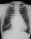
Figure 1
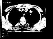
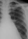
Figure 2
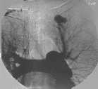
Figure 1 CXR shows a 2.5 cm mass in left apex with tubular structure connecting it to the left hilum. There is cardiomegaly with straight left heart border ,double atrial shadow and pulmonary venous hypertension typical of mitral valve disease.Figure 2 CXR localised view better demonstrates the mass to be connected by 2 tubular structure to the left hilum.Figure 3 CECT thorax shows the left apical mass to enhance to the same extent as adjacent vessels.Figure 4 IVDSA of thorax confirms the mass as a pulmonary AVM with a feeding artery and draining vein noted.
| ||||
HISTORY |
PREVIOUS MTHS |
|||
HOME |
CURRENT MTH |
|||