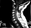
Figure 1

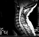
Figure 2
Figure 1 MRI(T1WI) shows a long spinal cord syrinx and the isointense tumour could not be clearly seen.Figure 2 MRI(PDWI) shows the syrinx to be hyperintense and again the mass is isointense.Figure 3 MRI (Gd-enhanced T1WI) demonstrates a strongly enhancing intramedullary mass in the upper cervical cord. Operative findings comfimed haemangioblastoma.
| ||||
HISTORY |
PREVIOUS MTHS |
|||
HOME |
CURRENT MTH |
|||