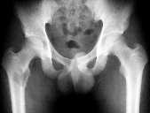
Figure 1
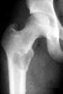
Figure 2
Figure 1 Pelvic x-ray shows a lucent lesion in proximal right femur, extending to the tip of lesser trochanter with cortical thinning and mild bony expansion seen.Figure 2 Right hip x-ray demontrates a narrow margin of transition with a punch out margin , minimal border sclerosis and slight periosteal reaction. Biopsy revealed Giant cell tumour.
| ||||
HISTORY |
PREVIOUS MTHS |
|||
HOME |
CURRENT MTH |
|||