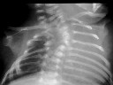
Figure 1
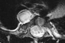
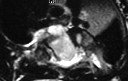
Figure 2
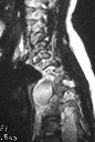
Figure 1 CXR shows segmentation abnormality in the mid-thoracic vertebrae.Figure 2-4 MR shows a right posterior mediastinal mass with CSF signal. It extends through the ventral defect of the thoracic vertebrae into the spinal canal. The thoracic cord is displaced to the right.
| ||||
HISTORY |
PREVIOUS MTHS |
|||
HOME |
CURRENT MTH |
|||