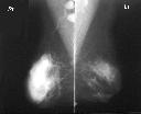
Figure 1
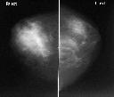
Figure 2
Figure 1 MLO view shows a large soft tissue mass(10 cm) in the right breast.The mass has a lobulated outline and partly indistinct border. Two enlarged lymph nodes with loss of hilar fat are seen in right axilla.Figure 2 CC view demonstrates the mass to have indistinct posterior border. Biopsy of the mass reviewed B cell lymphoma.
| ||||
HISTORY |
PREVIOUS MTHS |
|||
HOME |
CURRENT MTH |
|||