Figure 1
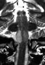
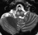
Figure 2
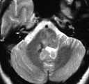
Figure 1 postoperative changes with hemosiderin deposition in the right dorsal pons and middle cerebellar peduncle.Figure 2 & 3 axial and coronal T2WI shows enlargement and increase signal intensity in bilateral inferior olive nuclei.
| ||||
HISTORY |
PREVIOUS MTHS |
|||
HOME |
CURRENT MTH |
|||