| ||||||||||||

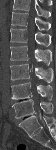
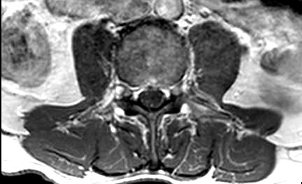

CLINICAL HISTORY: A middle age man presented with lower limb weakness |
||
PREVIOUS CASES |
||
HOME |
COMMENTS |
|
CASE OF THE MONTH |
|
|
|
||||||||||||||||||||||
|---|---|---|---|---|---|---|---|---|---|---|---|---|---|---|---|---|---|---|---|---|---|---|---|
Click on thumbnail for full-sized image
Disclaimer: The material used in this Web page is for educational purpose only.