| ||||||||
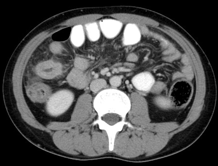
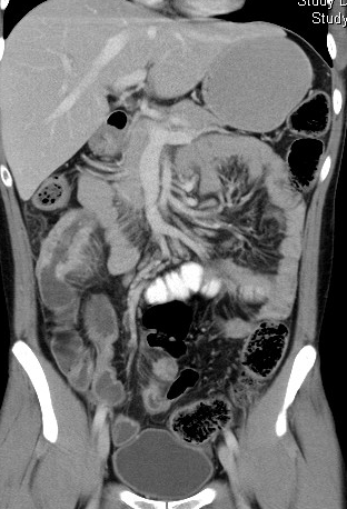

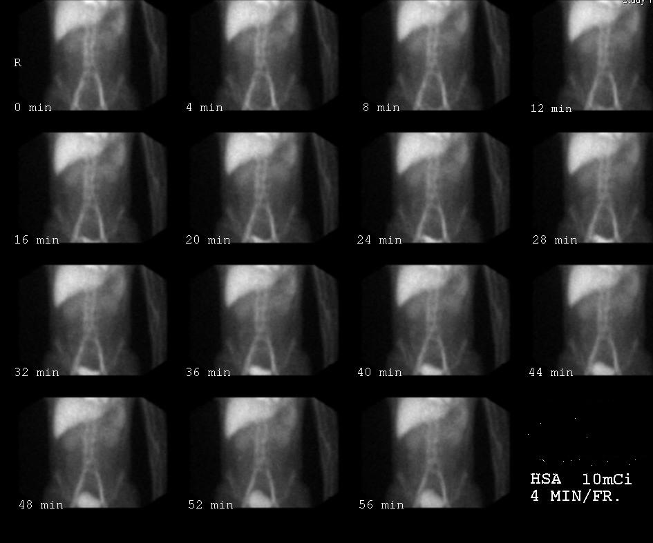
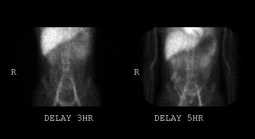
CLINICAL HISTORY: A 32-year-old man with known history of hypoalbuminaemia and H.pylori negative gastric ulcer presented with acute right lower quadrant pain and fever. CT abdomen was performed during acute presentation, human serum albumin scan was later performed. DIAGNOSIS: Crohn disease DISCUSSION: In young patients with acute right lower quadrant pain and fever, CT is often requested to look for evidence of acute appendicitis. In this set of CT, there was no significant inflammatory change of the caecum and appendix to suggest appendicitis. Multiple discontinuous segments of luminal narrowing and wall thickening were noted along the ileum. A segment of ileum in right mid-abdomen showed marked wall thickening and perifocal fat stranding, suggestive of inflammation. This inflamed segment appeared to be separated from the other bowel loops by the area of fat stranding, this was suggestive of creeping fat. Creeping fat and multiple discontinuous segments of bowel inflammation were typical of Crohn disease. Crohn disease can affect the entire GI tract, causing transmural inflammation, non-caseating granuloma, enlargement of submucosal lymphoid follicles, mucosal ulceration and fistula formation. Extraintestinal manifestations include liver abscess, gallstones, primary sclerosing cholangitis, urinary stones, bilateral sacroiliitis and erythema nodosum. |
||
PREVIOUS CASES |
||
HOME |
COMMENTS |
|