CLINICAL HISTORY:
A 34 year old lady with good past health complained of right submental swelling for 4 months. The lesion was non tender and soft on palpation. It did not move when swallowing or protruding tongue. Rest of the oral cavity was clear. MRI was performed for the lesion.
DIAGNOSIS:
Diving (Plunging) Ranula
DISCUSSION:
Ranula is mucus retention cyst arising from an obstructed sublingual or
minor salivary gland in the sublingual space. It usually presents as a
painless swelling during middle age. The lesion can be divided into simple
or diving types, with simple ranula confined to the sublingual space and
diving type herniating around or through mylohyoid muscle. Actually, diving
ranula is believed to result from rupture of simple ranula. When simple
ranula is left untreated, it will continue to grow and become ruptured,
dissecting down between facial planes into the neck either posterior to the
mylohyoid muscle or through a defect or vascular cleft in the mylohyoid
muscle.
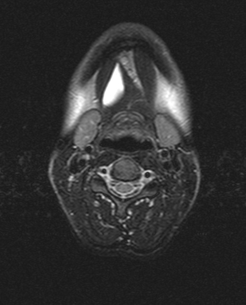
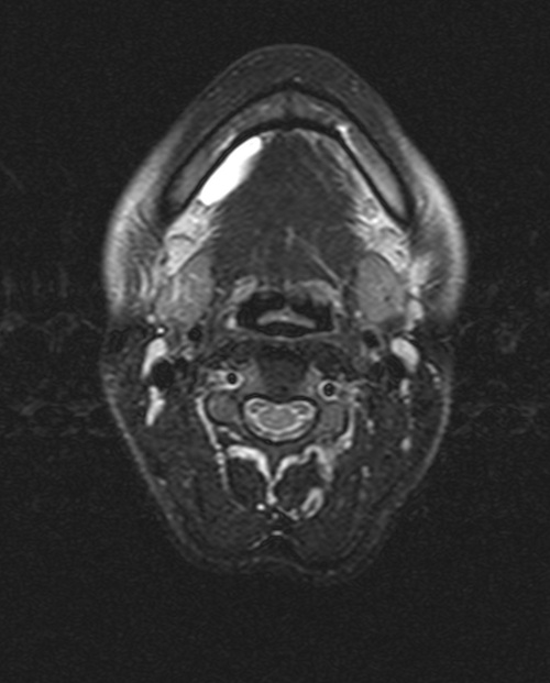
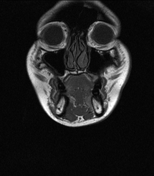
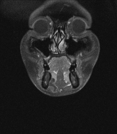
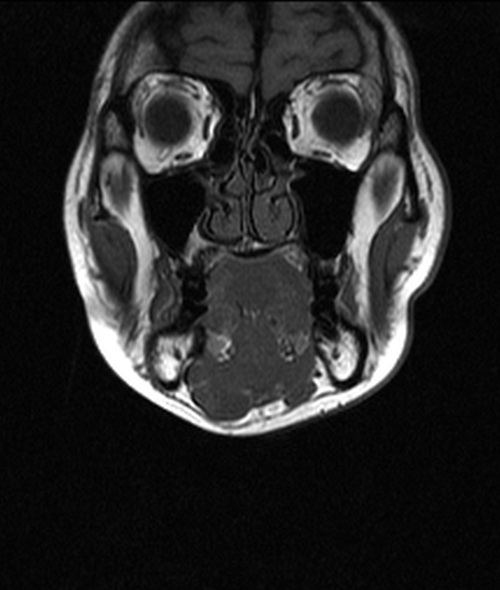
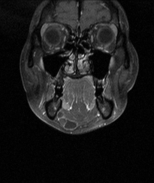
The ranula in our patient demonstrated the typical T1 hypointense and T2
hyperintense signal with thin rim enhancement. The coronal T1 post contrast
images with fat saturation revealed involvement of right sublingual space
and anterior aspect of right submandibular space. (Figures 4 and 6) It
suggested that the lesion in right sublingual space herniated through the
mylohyoid vascular cleft or defect into the anterior aspect of right
submandibular space, which is a less common route. For most ranula, the
lesion herniated directly posterior to the mylohyoid muscle into the
submandibular space.
Contrast-enhanced CT or MR images provide important information not only for
diagnosis of the disease but also differentiation of ranula from other
differenitial diagnoses, e.g. epidermoid cyst, dermoid cyst, brachial cleft
cyst. Imaging is also important in guiding surgery. Surgical excision of the
lesion is the treatment of choice, although some authors may believe
excision of psuedocyst is unnecessary. If the lesion is planned to be
excised, detailed mapping of the lesion provided by imaging can assist the
surgeon in planning the approach of resection. Complete resection of the
lesion is necessary to prevent recurrence of the lesion.
|