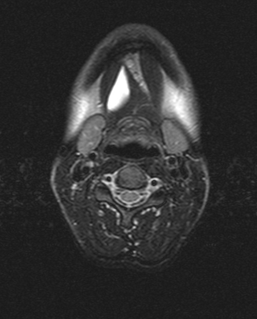
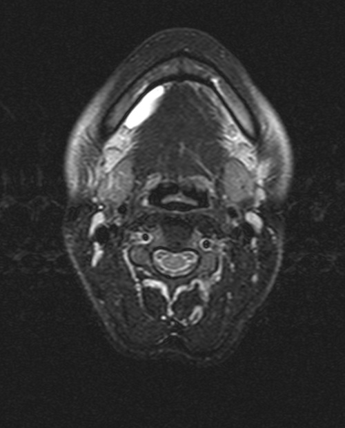
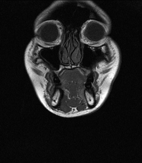
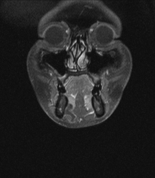
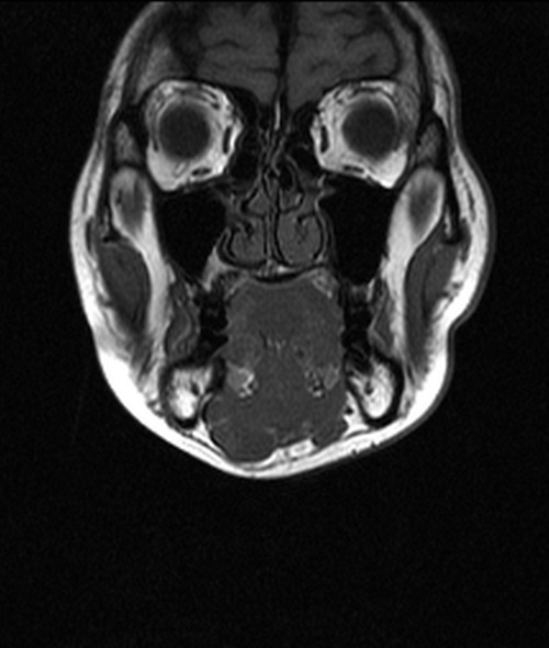
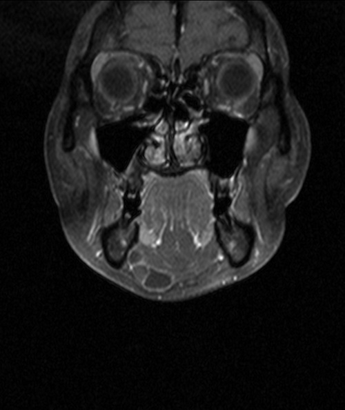
CASE OF THE MONTH

2012

November


Figure 1. T2 TSE axial MRI image with fat suppression of the floor of mouth


Figure 2. T2 TSE axial MRI image with fat suppression of the floor of mouth (cranial to Figure 1)


Figure 3. T1 SE Coronal MRI image of the floor of mouth


Figure 4. T1 SE Coronal MRI image with fat suppression and gadolinium contrast of the floor of mouth (Same level to Figure 3)


Figure 5. T1 SE Coronal MRI image of the floor of mouth (posterior to Figure 3)


Figure 6. T1 SE Coronal MRI image with fat suppression and gadolinium contrast of the floor of mouth (Same level to Figure 4)

CLINICAL HISTORY:

A 34 year old lady with good past health complained of right submental swelling for 4 months. The lesion was non tender and soft on palpation. It did not move when swallowing or protruding tongue. Rest of the oral cavity was clear. MRI was performed for the lesion.

PREVIOUS CASES

HOME

COMMENTS

Click on thumbnail for full-sized image


Disclaimer: The material used in this Web page is for educational purpose only.