|
Flair
|
Hemo
|
|
T1
|
T2
|
|
T1+C
|
|
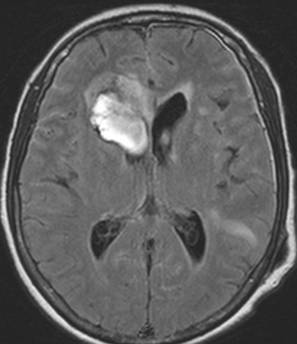
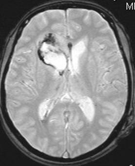
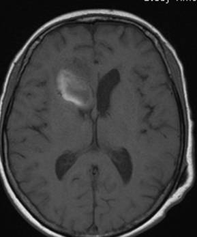
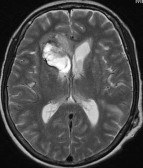
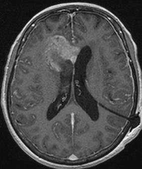
CLINICAL HISTORY: DIAGNOSIS: DISCUSSION: Glioblastoma multiforme is an aggressive tumor, it is the most common primary brain tumor in adults, accounting for 25% of all cases. It is also the most common tumor to involve the corpus callosum. Bihemispheric involvement by glioblastoma can result in a classical butterfly pattern. On MR imaging, these tumors typically show intense contrast enhancement. The MRI images also showed diffuse leptomeningeal metastasis with hyperintensity on FLAIR and enhancement on T1+C images. |
||
PREVIOUS CASES |
||
HOME |
COMMENTS |
|