|
|
|
|
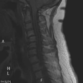
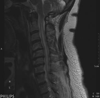
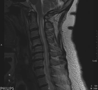
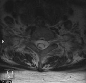
CLINICAL HISTORY: A 52-years-old lady complained of bilateral intrinsic hand muscles wasting for 10 yrs. Physical examination revealed weakness along bilateral C8 to T1 myotomes. Nerve conduction test showed normal sensory conduction. Plain MRI cervical spine (Sagittal T1W, Sagittal T2W, Axial T2W) was obtained. DIAGNOSIS : Chiari I malformation DISCUSSION: MRI cervical spine showed presence of hydrosyrinx along the spinal cord extending from C2 level downward. Associated expansion of the cord is discerned at the upper thoracic level with posterior scalloping of the adjacent vertebral bodies. Slight inferior herniation of the cerebellar tonsils is seen assuming a peg-like configuration. Overall features are compatible with Chiari I malformation. |
||
PREVIOUS CASES |
||
HOME |
COMMENTS |
|