|
CLINICAL HISTORY:
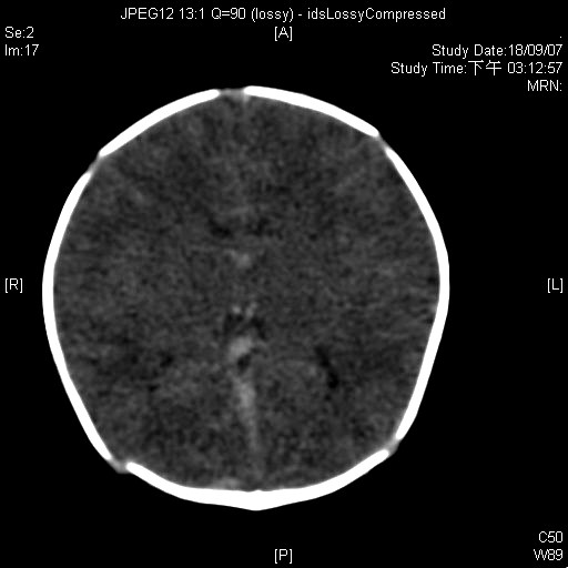
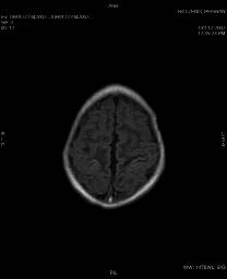
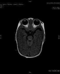
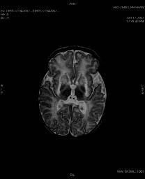
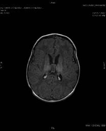
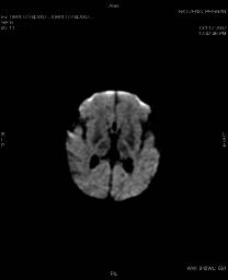
A term newborn boy was delivered by Caesarean Section at 38th week of gestation. The baby's mother was 16 years old and lived in China but antenatal care was performed in HK and was uneventful. No history of maternal drug abuse or maternal fever. The baby was found to be flaccid and apneic at delivery and required active resuscitation and he was intubated. The Agar Score was 1,4,6. On arrival to nursery at about 30 min, gasping effort was noted but there was no regular breathing effort and muscle tone increased. H'stix was 10.9. First venous blood gas showed pH of 7.3. CT scan and MRI scan were performed on Day 2 and 1 month later respectively.
DIAGNOSIS
Hypoxic ischemic encephalopathy
DISCUSSION
MRI is the most sensitive and specific imaging modality for the diagnosis of hypoxic-ischemic brain injury. Hypoxic-ischemic injury to gray matter (deep gray matter, cortex) demonstrates characteristic T1 hyperintensity and variable T2 intensity, depending on the time at imaging and the dominant underlying pathologic condition, such as hemorrhage or gliosis. Injury to white matter generally results in T1 hypointensity and T2 hyperintensity due to ischemia-induced edema. Diffusion-weighted MR imaging performed with apparent diffusion coefficient maps between 24 hours and 8 days of life is more sensitive for the detection of cytotoxic edema, as it reveals restricted diffusion earlier than the signal intensity abnormalities evident on conventional T1- or T2-weighted images. However, the apparent diffusion coefficient value does not appear to correlate well with the extent of the ischemic injury and is not predictive of adverse outcome. MR spectroscopy provides gross biochemical analysis of the "compromised anaerobic" cerebral tissues, as it reveals changes in the concentrations of lactate, choline, creatine, N-acetylaspartate (NAA), and glutamine. MR imaging is also helpful in excluding other causes of encephalopathy such as hemorrhage, cerebral infarction, neoplasms, or congenital malformations. |