|
Clinical History
A 38 year-old man with otherwise good past health presented with headache.
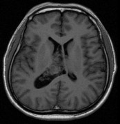
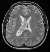
Diagnosis: Central Neurocytoma
Comment:
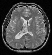
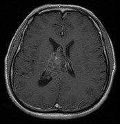
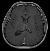
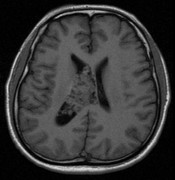
The lesion is T1W isointense and T2W hyperintense. It situates within the body of right lateral ventricle. It is attached to the septum pellucidum which is displaced to the left side. The tumour shows characteristic bubbly appearance with multiple small cystic components which are T1W hypointense and T2W hyperintense. Mild contrast enhancement is present. The right lateral ventricle is mildly dilated due to obstruction of the interventricular Foramen of Monro.
Central neurocytoma is most commonly found at the body or frontal horn of lateral ventricle and 15% shows extension into the third ventricle. They are rarely found in the brain parenchyma or spinal cord. Calcification is depicted in 50 to70 % of cases on CT scan. They are rarely complicated by hemorrhage. Extraventricular extension of this tumour signifies poorer prognosis.
|