|
Clinical History
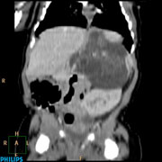
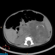
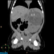
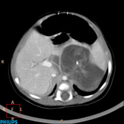
59-day-old baby girl presented with antenatal USG finding of polycystic mass in left renal region.
Diagnosis: Mature Teratoma
Comment :
The radiological features supporting the diagnosis of mature teratoma include calcification in linear strand pattern in the noncontrast coronal view which resembles architecture of osseous component. In neuroblastoma, it is usually associated with regular stippled calcification rather than linear calcification. In addition, from the postcontrast axial and coronal view, this mass displaces adjacent SMA and celiac trunk rather than encasing them. This again favours the diagnosis of mature teratoma.
|