CASE OF THE MONTH

2007

May


Figure 1. USG of the Urinary Bladder (Transverse Scan)


Figure 2. USG of Urinary Bladder (Longitudinal Scan)


Figure 3. CT scan of the Pelvis

Figure4
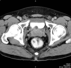
Figure 4. CT scan of the pelvis in delayed phase.

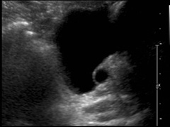
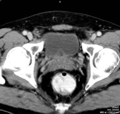
Clinical History

A 54 year old gentleman with hypercholesterolaemia.

Diagnosis:  Mullerian prostatic cyst.

Comment :
Ultrasound shows a well defined smooth roundish unilocular cystic lesion seen in bladder base in midline. No colour Doppler flow signal is seen within this lesion. Prostate is not enlarged. CT shows a cystic lesion indenting on the bladder base, likely extra-vesical and arising from the anterosuperior aspect of midline of prostate.
Mullerian prostatic cyst is a congenital midline cystic structure which is developmentally derived from the Mullerian ducts. A paramedian location has been reported. The cyst usually extends above the base of the prostate.On either endorectal ultrasound, CT or MRI, a midline or paramedian cystic structure, extending above the base of the prostate gland, is virtually pathognomonic.

PREVIOUS CASES

HOME

COMMENTS

Click on thumbnail for full-sized image


Disclaimer: The material used in this Web page is for educational purpose only.