Clinical History
A 56-year-old lady with a known residual right
pontine cavernous malformation since 2002 (excision done in 2004)
presented with persistent ataxia. These are selected images from
the follow-up MRI performed in 2006.
Diagnosis:
Hypertrophic olivary degeneration
Comment :
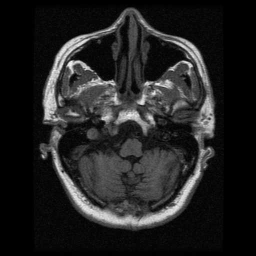
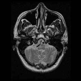
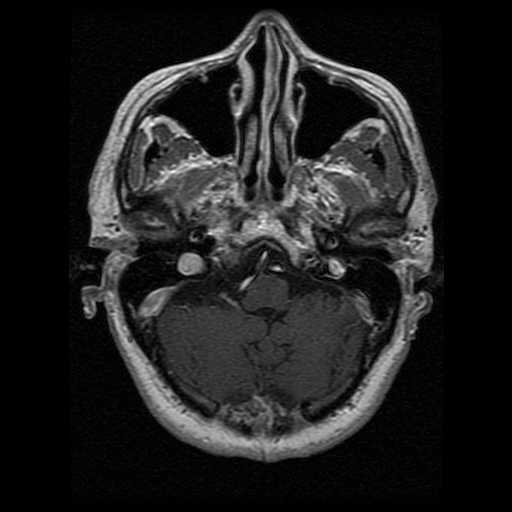
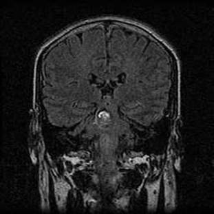
On the coronal FLAIR image, the heterogeneously hyper- to
isointense mass lesion in the right side of the pons represents the residual
pontine cavernous haemangioma. A rim of haemosiderin deposition is also
evident. On the T2W axial and the coronal FLAIR images, the right inferior
olivary nucleus is enlarged and shows increased signal intensity. It
appears isointense on T1WI and is non-enhancing following contrast
administration. The overall features are those of hypertrophic olivary
degeneration. Hypertrophic olivary degeneration is a form of transneuronal
degeneration caused by lesions in the Guillain-Mollaret triangle. This
circuit connects the inferior olivary nucleus (ION), red nucleus (RN) and
contralateral dentate nucleus (DN) . |