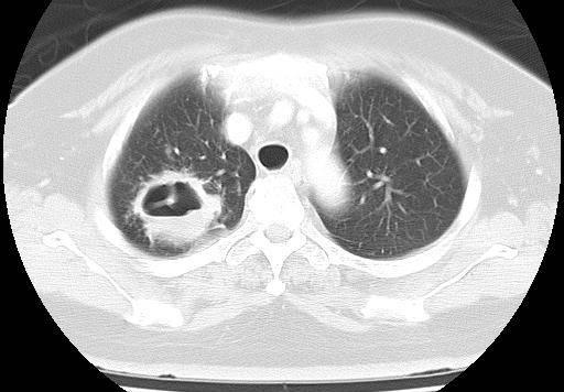
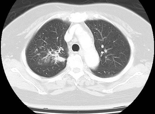
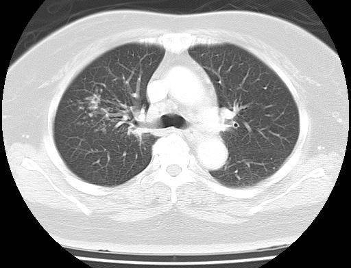
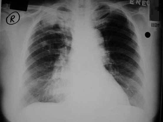
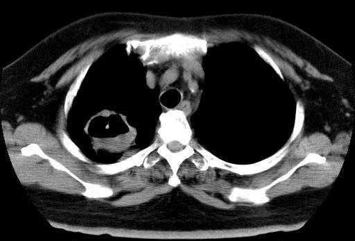
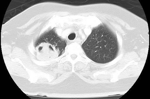
CASE OF THE MONTH

2006

Sep


Clinical History

A 62 years old lady with history of HT and DM, admitted for malaise, weight loss and cough. CXR and CT thorax was done.

Diagnosis:

Tuberculosis

Comment:

Plain chest radiograph shows a cavitating lesion over the right upper zone,
overlapping the posterior right 5th rib. There is also consolidation of the
right middle lobe, with blurring of the right heart border. Computed
tomography (CT) scan confirms the presence of a cavitating lesion over the
right upper lobe, with air-fluid level demonstrated. Centrilobular nodules
are seen in the lung window. Features are suggestive of post-primary
pulmonary tuberculosis.


PREVIOUS CASES

HOME

COMMENTS

Click on thumbnail for full-sized image


Disclaimer: The material used in this Web page is for educational purpose only.