Diagnosis:
Vertebral artery dissection
Comment :
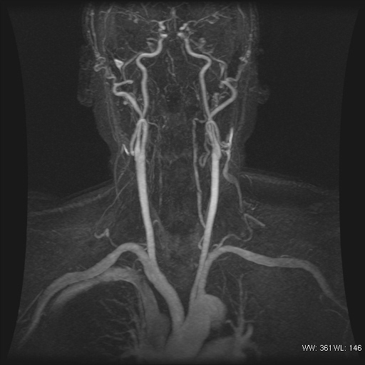
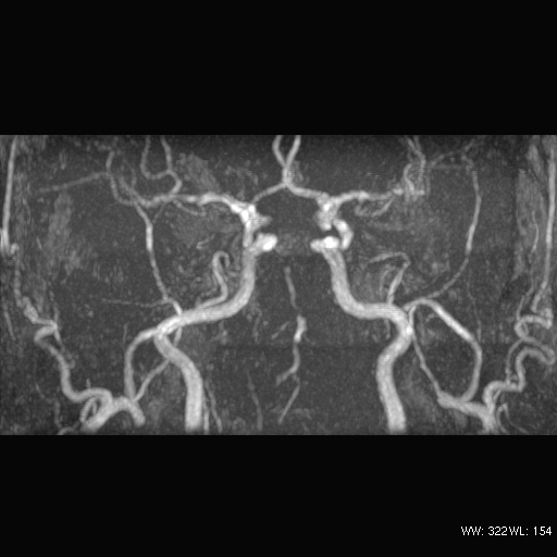
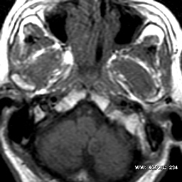
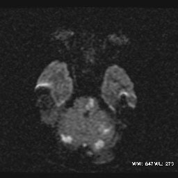
The imaging findings are that of steno-occlusive pattern
of vertebral artery dissection. The contrast enhanced MR anigogram
clearly shows the tapered occlusion of the vertebral arteries at
V3 levels. V2 segments also show irregular filling defects. Very
little reconstitution of intracranial vertebral and basilar arteries
can be seen. Also noted the high signal in the left intracranial
vertebral artery on T1W axial MR image, probably due to subacute
clot inside or very slow flow. T1W axial scan of the neck failed
due to patient movement despite sedation. The classical finding
would be a crescent of high signal intramural haematoma.
|