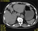
Figure 1: Noncontrast CT scan of liver.
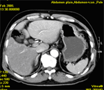
Figure 2: Contrast CT scan of liver during arterial phase.
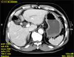
Figure 3: Contrast CT scan of liver during portovenous phase.
Clinical History72 years old male, presents with deranged liver function tests.Diagnosis:Transient hepatic attenuation difference secondary to thrombosis of left branch of portal vein Comment : Figure 1 : There is a curvilinear dense opacity in left lobe of liver. Figure 2 : Intense enhancement of left lobe of liver compared with right lobe is seen. Figure 3 : There is homogeneous enhancement of both lobes of liver.. |
||
PREVIOUS CASES |
||
HOME |
COMMENTS |
|