Fig.1
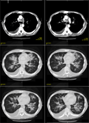
Diagnosis : Kaposiˇ¦s sarcoma (KS).Findings :CT scan shows the typical features of pulmonary KS with irregular and ill-defined peribronchovascular flame-shaped nodules and peribronchovascular interstitial thickening in both lungs. There are also interlobular septal thickening, pleural effusions, and mediastinal lymphadenopathy.88% of HIV-infected patient who showed peribronchovascular distribution of nodules had KS. This finding, in association with nodule size larger than 1cm, strongly predicts KS. |
||
DIAGNOSIS : |
Pulmonary varix t |
|
PREVIOUS CASES |
||
HOME |
COMMENTS |
|