CASE OF THE MONTH

2003

ANSWER

FOR Nov


Fig 1
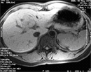
Precontrast T1-weighted

Fig 2
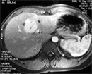
Postcontrast Arterial phase T1-weighted

Fig 3
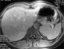
Postcontrast Delayed phase T1-weighted

  • A 3 cm well-defined right paracardiac opacity is seen on the frontal CXR. The opacity is situated in the right lower lobe, as it is posterior to the major fissure on the lateral view.
  • The lesion is demonstrated as a dilated right inferior pulmonary vein, draining into the left atrium. It shows comparable contrast enhancement to the left atrium.

DIAGNOSIS :

Pulmonary varix t

PREVIOUS CASES

HOME

COMMENTS

Click on thumbnail for full-sized image


Disclaimer: The material used in this Web page is for educational purpose only.