Figure 1
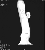
Figure 2
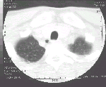
Figure 1: Thin sections CT thorax- An oval airspace is seen in the right posterolateral aspect of trachea at level of sternal notch.Figure 2: 3D reconstruction shows a neck between the airspace and trachea. |
||
DIAGNOSIS : |
Tracheal DiverticulumNote: The prevalence of tracheal diverticulum was reported to be about 1% in an autopsy series by Mackinnon. Tracheal diverticulum has also been reported to have an association with chronic obstructive airway disease. Presenting features include prolonged productive cough, hemoptysis and chest pain. |
|
PREVIOUS CASES |
||
HOME |
COMMENTS |
|